Thinking about robotic knee replacement in Navi Mumbai? It’s a valid question, and the short answer is yes, it’s a real option here, offering a potentially more precise approach compared to traditional surgery. This technology is becoming more accessible, and several hospitals in Navi Mumbai have adopted it. Let’s dive into what that actually means for you, the practicalities, and what to expect.
Robotic assistance in knee replacement surgery isn’t about a robot performing the entire operation autonomously. Instead, the robot acts as an advanced tool guided by the surgeon. It uses pre-operative imaging and intra-operative feedback to help the surgeon execute the procedure with enhanced precision.
How the Robot Assists
The robotic system typically involves a robotic arm that the surgeon directly controls. Before surgery, detailed scans (like CT scans) of your knee are used to create a 3D model. This allows the surgeon to plan the surgery meticulously, including the exact size and placement of the implants.
Key Benefits of Robotic Systems
The main advantage lies in precision. The robotic arm can help the surgeon make very accurate bone cuts, place the implant components in the ideal position, and balance the ligaments around the new knee. This level of accuracy can lead to better alignment and potentially a more natural feel for the replaced knee.
Not a “Robot Surgeon”
It’s crucial to understand that the surgeon is always in control. The robot is an extension of their hand, providing a stable platform and guided precision. It doesn’t make decisions; it executes the surgeon’s commands with exceptional accuracy.
Robotic knee replacement surgery has gained significant attention in Navi Mumbai for its precision and improved recovery times. For those interested in learning more about advanced orthopedic treatments, a related article can be found at Orthopedic Care in Thane, which discusses the latest innovations in joint replacement procedures and highlights the expertise of leading orthopedic surgeons in the region. This resource provides valuable insights into the benefits of robotic-assisted surgeries and how they are transforming patient outcomes.
Robotic Knee Replacement in Navi Mumbai: Where to Find It
Navi Mumbai has a growing number of healthcare facilities that have invested in this advanced surgical technology. You’ll find robotic systems available in some of the larger, multi-specialty hospitals.
Leading Hospitals Offering Robotic Surgery
Several prominent hospitals in the Navi Mumbai region have introduced robotic knee replacement programs. These institutions often house experienced orthopedic surgeons, many of whom have received specialized training in robotic-assisted procedures.
Identifying Hospitals with Robotic Capabilities
When researching, look for hospitals that explicitly mention robotic-assisted orthopedic surgery or specifically robotic knee replacement on their websites or through direct inquiry. Don’t hesitate to call and ask about their robotic platforms and the surgeons who utilize them.
The Role of Surgeon Experience
While the technology is important, the surgeon’s expertise remains paramount. A skilled surgeon, even with traditional methods, can achieve excellent results. However, combining a skilled surgeon with robotic assistance can potentially elevate the precision of the procedure further.
Training and Specialization in Robotic Surgery
Surgeons who use robotic systems undergo specific training for that particular platform. This training focuses on the nuances of controlling the robotic arm, interpreting the pre-operative plan, and integrating the robotic guidance into their surgical workflow.
The Surgical Process with Robotic Assistance

The steps involved in a robotic knee replacement are largely similar to traditional surgery, but with enhanced guidance during critical phases.
Pre-Operative Planning is Crucial
Before surgery, a CT scan of your knee is taken. This data is uploaded into the robotic system, which then creates a detailed 3D model. Your surgeon uses this model to map out the precise cuts and implant positioning, taking into account your unique anatomy.
Customizing the Surgical Plan
This pre-operative planning allows for a highly personalized approach. The surgeon can simulate different implant sizes and orientations to achieve the best possible outcome for your specific knee.
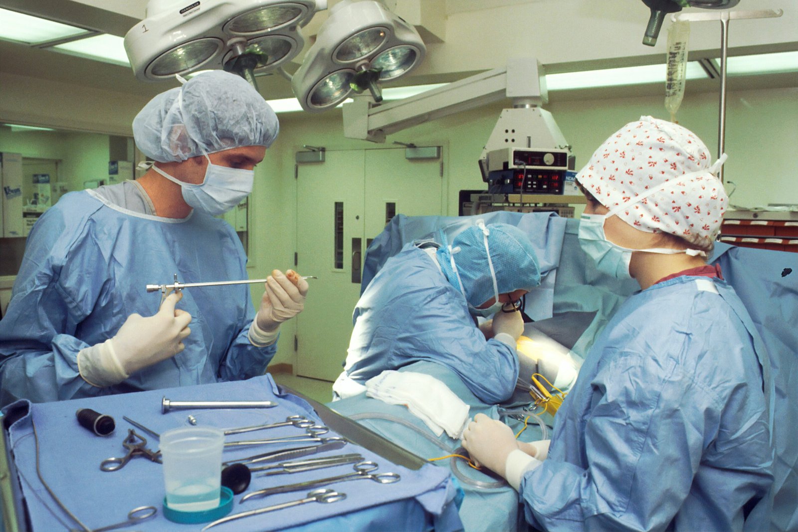
During the Surgery
Once you’re under anesthesia, the surgeon prepares the knee for the procedure. The robotic arm is then brought into the surgical field and the instruments are attached to it. The surgeon uses a console to control the robotic arm, which guides the instruments for bone preparation and implant placement.
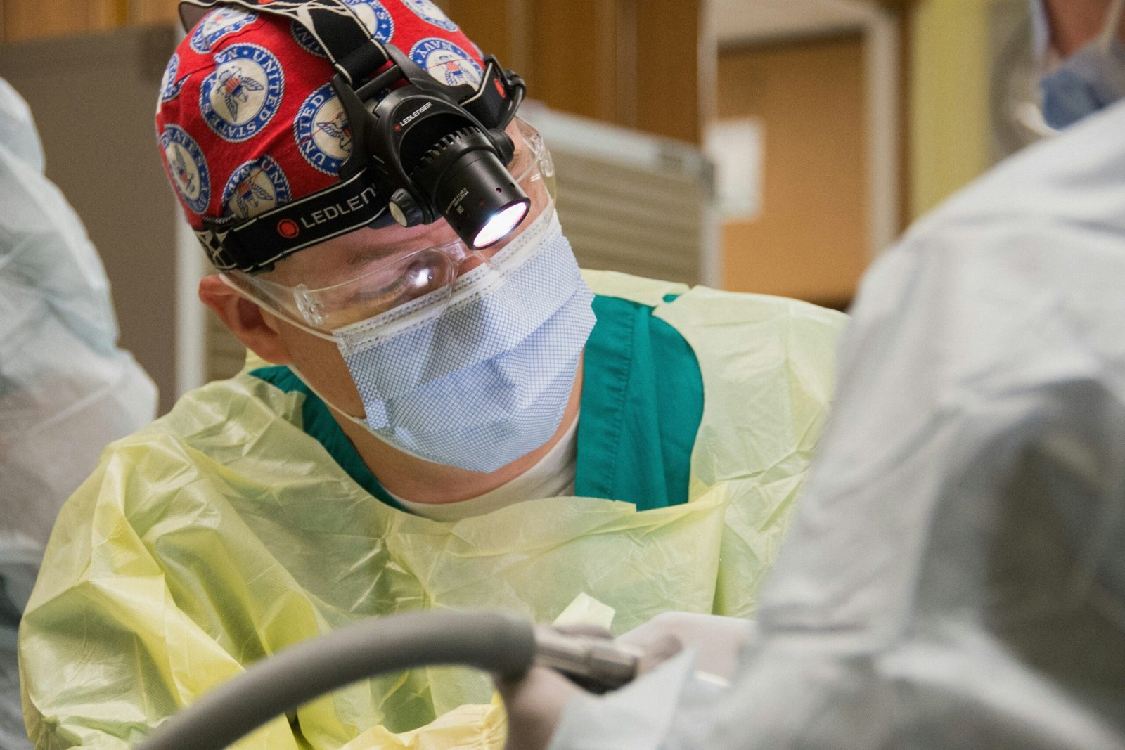
Interacting with the Robotic System
The surgeon essentially “docks” surgical instruments to the robotic arm. They then use hand controls and a visual interface to guide the instruments. The system provides real-time feedback, and importantly, has built-in safety features that prevent the robotic arm from moving outside the pre-defined surgical plan.
Implant Placement and Ligament Balancing
The robot helps achieve extremely accurate bone cuts. This allows for precise placement of the prosthetic components. Equally important is ligament balancing, ensuring the knee has proper stability and range of motion after the surgery. Robotic systems can assist in achieving optimal tension in the ligaments.
Ensuring Accurate Implant Alignment
Correct alignment of the implant is vital for long-term success and minimizing wear. The robotic system’s precision in bone preparation and implant placement can contribute to better alignment.
Recovery and Potential Advantages of Robotic Assistance

While the fundamental principles of recovery remain the same, some studies suggest potential advantages with robotic-assisted procedures.
Post-Operative Recovery Trends
Many patients report a smoother recovery, potentially related to the heightened precision of the surgery. This might translate to reduced pain in the initial days and a quicker return to certain activities. However, individual recovery varies greatly.
Pain Management and Rehabilitation
Pain management and adherence to your rehabilitation program are still the cornerstones of recovery, regardless of the surgical technique used. Your physiotherapy will be tailored to your specific needs.
Potential for Improved Outcomes
The goal of robotic assistance is to achieve more predictable and consistent results. Some research indicates that robotic-assisted knee replacements may lead to better implant longevity and patient satisfaction due to improved alignment and fit of the implants.
Long-term Durability and Function
By enabling more accurate placement and balancing, the hope is that robotic surgery can contribute to the long-term durability and optimal function of the artificial knee joint.
Faster Return to Activities?
While not guaranteed, some individuals may experience a slightly quicker return to daily activities. This is often attributed to reduced tissue trauma during surgery and the potential for improved knee mechanics right from the start.
Robotic knee replacement surgery has been gaining popularity in Navi Mumbai due to its precision and faster recovery times. Patients seeking advanced treatment options can find valuable insights in a related article that discusses the benefits and advancements in this innovative surgical technique. For more information, you can read about the professional staff involved in these procedures by visiting this link. The expertise of the medical team plays a crucial role in ensuring successful outcomes for patients undergoing robotic knee replacements.
Choosing the Right Surgeon and Hospital in Navi Mumbai
| Hospital | Success Rate | Recovery Time | Cost |
|---|---|---|---|
| Hiranandani Hospital | 95% | 2-3 weeks | INR 3,50,000 |
| Fortis Hospital | 92% | 3-4 weeks | INR 3,00,000 |
| Reliance Hospital | 90% | 2-3 weeks | INR 3,20,000 |
Selecting your surgeon and hospital is a critical step, whether you opt for robotic or traditional knee replacement.
Researching Orthopedic Surgeons
Look for orthopedic surgeons who specialize in knee replacement and have experience with robotic-assisted procedures. Don’t be afraid to ask about their training, the number of robotic surgeries they perform, and their patient outcomes.
Dr. [Your Surgeon’s Name]’s Approach
(This is where you’d slot in a specific surgeon if you were writing a factual piece with named examples. For this general article, we’ll keep it generic.) A good surgeon will clearly explain the procedure, discuss the pros and cons of different approaches, and answer all your questions thoroughly.
Hospital Facilities and Technology
Ensure the hospital you choose is well-equipped to handle complex orthopedic surgeries and has the specific robotic platform you and your surgeon decide is best.
Evaluating Hospital Accreditations and Staff
Check the hospital’s accreditations and the experience of their nursing and support staff. A supportive environment contributes significantly to a positive recovery experience.
Cost Considerations
Robotic knee replacement surgery can sometimes be more expensive than traditional surgery due to the cost of the technology. It’s important to discuss the total cost, including implants, hospital stay, and physiotherapy, with the hospital and your insurance provider.
Understanding Insurance Coverage
Verify what your health insurance policy covers for robotic-assisted surgeries. Many policies cover the procedure itself, but you’ll need to confirm any specific limitations or co-pays related to the robotic component.
In recent advancements in orthopedic procedures, robotic knee replacement surgery has gained significant attention in Navi Mumbai for its precision and improved recovery times. A related article discusses the benefits of pediatric orthopedic surgery, highlighting how innovative techniques can enhance treatment outcomes for younger patients. For more information, you can explore the article on pediatric orthopedic surgery here. This connection underscores the importance of technological advancements in various age groups, ensuring better care and rehabilitation for all.
Frequently Asked Questions About Robotic Knee Replacement
Here are some common queries people have when considering this surgical option.
Is Robotic Surgery More Painful?
Generally, the aim of robotic surgery is to minimize disruption, which can potentially lead to less post-operative pain. However, pain levels are highly individual and depend on many factors.
Pain Management Strategies
Your medical team will have a comprehensive pain management plan in place to help you through your recovery.
What is the Recovery Time?
While recovery timelines vary, some individuals might find their initial recovery slightly quicker with robotic assistance. However, a full return to all activities typically takes several months, with consistent physiotherapy being essential.
The Importance of Physiotherapy
Your rehabilitation program is crucial for regaining strength, flexibility, and function. Do not skip your physiotherapy sessions.
Am I a Candidate for Robotic Knee Replacement?
This is a decision best made in consultation with your orthopedic surgeon. They will assess your knee condition, overall health, and lifestyle to determine if robotic surgery is the most suitable option for you.
Surgeon’s Recommendation is Key
Ultimately, your surgeon’s recommendation, based on their expertise and understanding of the robotic system, will guide your decision.
What are the Risks?
As with any surgery, there are risks involved, including infection, blood clots, and implant-related issues. The use of robotic technology aims to minimize some surgical risks through enhanced precision, but it doesn’t eliminate all potential complications.
Discussing Risks with Your Surgeon
It’s vital to have an open and detailed discussion with your surgeon about the specific risks and benefits of robotic knee replacement in your case.
What if the Robot Malfunctions?
Robotic surgical systems are designed with multiple fail-safes. If there were any technical issues, the surgeon would revert to traditional manual techniques, ensuring the surgery can proceed safely. The surgeon remains in complete control throughout the procedure.
FAQs
What is robotic knee replacement surgery?
Robotic knee replacement surgery is a minimally invasive procedure that uses a robotic arm to assist the surgeon in performing the surgery with a high level of precision. It allows for more accurate placement of the knee implant, resulting in better outcomes for the patient.
How is robotic knee replacement surgery performed?
During robotic knee replacement surgery, the surgeon uses a robotic arm to create a 3D model of the patient’s knee and then uses this model to plan and execute the surgery with a high level of accuracy. The robotic arm assists the surgeon in removing damaged bone and cartilage and in placing the knee implant in the optimal position.
What are the benefits of robotic knee replacement surgery?
Robotic knee replacement surgery offers several benefits, including smaller incisions, less damage to surrounding tissue, reduced pain and scarring, faster recovery time, and improved long-term outcomes for the patient. The high level of precision provided by the robotic arm also leads to better alignment and positioning of the knee implant.
Who is a candidate for robotic knee replacement surgery?
Candidates for robotic knee replacement surgery are typically individuals with severe knee pain and disability caused by conditions such as osteoarthritis, rheumatoid arthritis, or post-traumatic arthritis. The decision to undergo robotic knee replacement surgery is made on a case-by-case basis after a thorough evaluation by a surgeon.
Where can I undergo robotic knee replacement surgery in Navi Mumbai?
Robotic knee replacement surgery is offered at various hospitals and medical centers in Navi Mumbai. Patients can consult with orthopedic surgeons specializing in robotic surgery to determine the best course of treatment for their specific condition.